If you are interested in understanding the structure and functioning of the human respiratory system, you must read on; for this Bodytomy article provides you with information about the same with images.
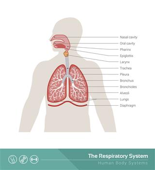
The human respiratory system is composed of the nasal passages, the pharynx, larynx, the trachea, bronchi, and the lungs. It is responsible for the process of respiration that is vital to the survival of living beings. Respiration is the process of obtaining and using oxygen, while eliminating carbon dioxide. It is the process by which human beings take in the oxygen from their environment and give out carbon dioxide that is produced as a result of chemical reactions within the cells. The human respiratory system is the specialized system that brings about this critical process in human beings. Let us look at its structure.
The respiratory system in human beings can be divided into the upper respiratory tract that consists of nasal passages, pharynx, and larynx, and the lower respiratory tract that is composed of the trachea, the primary bronchi, and the lungs. This Bodytomy article tells you about the human respiratory system structure with the help of diagrams.
Nasal Cavity
Air entering from the nostrils is led to the nasal passages. The nasal cavity that is located behind the nose comprises the nasal passages that form an important part of the respiratory system in human beings. The nasal cavity is responsible for conditioning the air that is received by the nose. The process of conditioning involves warming or cooling of the air received by the nose, removing dust particles from it, and also moistening it before it enters the pharynx.

Human Respiratory System
Pharynx
It is located behind the nasal cavity and above the larynx. It is also a part of the digestive system. Food as well as air pass through the pharynx.

Pharynx Structure
The upper part of the pharynx is called nasopharynx. It is located above the oral cavity, extending from the base of the skull to the upper surface of the soft palate. Located in its posterior wall are adenoids or pharyngeal tonsils. Pharyngeal ostia are located on its lateral walls. Lying behind the oral cavity is the oropharynx. It extends from the uvula up to the hyoid bone and opens into the mouth. Located on its anterior wall are the base of the tongue and the epiglottic vallecula. The tonsil, tonsillar fossa, and tonsillar pillars are located on its lateral wall. The inferior surface of the soft palate and the uvula are located on its superior wall, while the palatine tonsil is located on the lateral wall of the oropharynx. The part of the throat that connects to the esophagus is the laryngopharynx. Located below the epiglottis, it extends to the point where this common pathway diverges into two; the respiratory pathway and the digestive pathway. Food and air pass through the laryngopharynx.
Larynx
It is associated with the production of sound. It consists of two pairs of membranes. Air causes the vocal cords to vibrate, thus producing sound. The larynx is situated in the neck of mammals and plays a vital role in the protection of trachea.

Larynx Structure
Supporting the mammalian larynx are nine cartilages. The thyroid cartilage forms the Adam’s apple. The inferior wall of the larynx is formed by cricoid cartilage attached to the top of the trachea. A large piece of elastic cartilage forms the epiglottis. When the larynx elevates, it moves down to form a cover over the glottis, thus closing it. The arytenoid cartilages affect the position and tension of the vocal folds. Located on top of these are the corniculate cartilages. Positioned anterior to these are the cuneiform cartilages.
The intrinsic muscles of the larynx are classified as respiratory and phonatory. The former enable breathing while the latter are responsible for production of sound. The extrinsic muscles support and position the larynx.
Trachea
This refers to the airway through which respiratory air travels. The rings of cartilage within its walls keep the trachea open. It connects the larynx and the pharynx to the lungs.

Trachea Anatomy
Around 1 inch in diameter and 10 to 16 cm long, it extends from the base of the larynx to the level of thoracic vertebra T5. The airway is protected by incomplete C-shaped rings of cartilage. The trachealis muscle connects the ends of these rings. Located towards the rear of the trachea is the esophagus. The epiglottis closes the opening to the larynx, so as to not let the swallowed matter enter the trachea.
Bronchi
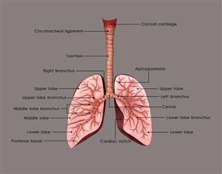
The trachea are divided into two main bronchi. The bronchi extend into the lungs spreading in a tree-like manner as bronchial tubes. The bronchial tubes subdivide and with each subdivision, their walls get thinner. This dividing of the bronchi into thin-walled tubes results in the formation of bronchioles. The bronchioles terminate in small air chambers, each of which contains cavities known as alveoli. Alveoli have thin walls which form the respiratory surface. The exchange of gases between the blood and the air takes place through these walls.
The right main bronchus enters the right lung. It divides into three secondary bronchi that supply air to the superior, middle, and inferior lobe of the right lung. The left main bronchus enters the root of the left lung. It divides into two secondary bronchi that supply air to the superior and inferior lobes of the left lung. The secondary bronchi divide into tertiary bronchi, which further divide to form bronchioles. These smaller bronchioles divide into terminal bronchioles, that further form respiratory bronchioles. They divide into alveolar ducts, each of which has 5-6 alveolar sacs associated with it.
Bronchioles
They refer to the ways through which air passes through the nose or mouth to the alveoli. Usually, bronchioles are less than 1 mm in diameter. They are supported by elastic fibers attached to the surrounding lung tissue.
Alveoli
They are located where the alveolar ducts and atria terminate. They are wrapped in capillaries. They contain elastic fiber and collagen. The elastic fibers enable their stretching during inhalation of air.
Lungs
Lungs form the most vital component of the human respiratory system. They are located on the two sides of the heart. They are responsible for transporting oxygen into blood and releasing carbon dioxide from it. The two lungs are not identical. The right lung consists of three lobes while the left one consists of two. The right lung has an almost vertical medial border. The left lung has a cardiac notch that holds the heart. The lobes of the lungs have pleural cavities. The cavity helps lubricate the lungs and also helps the lung surface remain in contact with the rib cage.
Diaphragm
Separating the thoracic cavity from the abdominal cavity, the diaphragm is located at the bottom of the rib cage, and made up of a sheet of internal skeletal muscle and fibrous tissue. It is in the shape of a dome, with its upper surface forming the base of the thoracic cavity and its lower surface forming the roof of the abdominal cavity. When air is inhaled, the diaphragm contracts and moves downwards to expand the thoracic cavity. When the diaphragm relaxes, the lung and tissues that line the thoracic cavity recoil, and air is expelled.
This was a brief description of the human respiratory system structure. It is that vital system in our body, which enables us to literally ‘breathe new life’ every instant.
