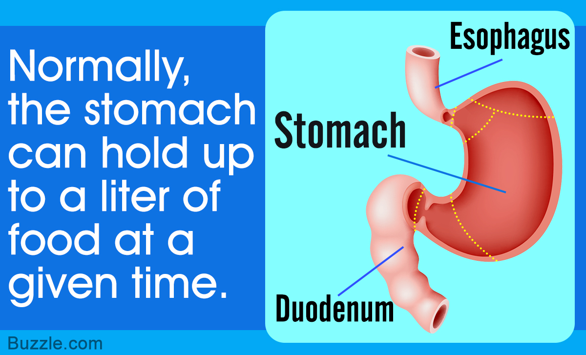
A hollow, J-shaped muscular organ, the stomach is an important part of the digestive system of the human body. Bodytomy provides information on the location and functions of the stomach, along with a labeled diagram to help you understand the anatomy of the human stomach.
Did You Know?
To provide protection from the highly acidic environment, the epithelial cells that line the stomach produce a new layer of mucus every two weeks.
Located in the upper-left section of the abdomen, the stomach is a visceral organ that lies beneath the diaphragm. The average length of this muscular organ is about 10 inches. It extends between the seventh thoracic vertebra (T7) and the third lumbar vertebra (L3). It is an organ of the digestive system. When we eat, enzymes in the mouth act on the food. This food then passes from the esophagus to the stomach. The stomach acts as a storage vessel for the food. Normally, the stomach can hold up to a liter of food at a given time. This capacity can vary from person to person.
Food usually remains in the stomach for about two hours. During this time, the gastric enzymes act on the food, turning it into chyme, which refers to partially-digested, semi-liquid mass of food. By peristalsis or the wave-like muscle contractions, the partially-digested food is moved from the stomach into the small intestine, where most of digestion and absorption of nutrients takes place.
Anatomy of the Stomach
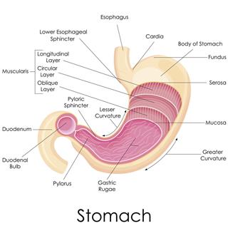
The human stomach has two surfaces, two orifices, and two curvatures. The two orifices of the stomach are called cardiac orifice and pyloric orifice. The former is located on the left, at the level of the tenth thoracic vertebra (T10). The pyloric orifice lies at the level of the first lumbar vertebra (L1). It is the opening from where the food travels towards the duodenum. Given below is a labeled diagram of the stomach to help you understand stomach anatomy.
The stomach is divided into four parts. These include:
- Cardia
- Fundus
- Body
- Pylorus
Cardia refers to the section of the stomach that is located around the cardiac orifice. The lower esophageal sphincter lies at the junction where the esophagus meets the stomach. It prevents the backflow of food from the stomach to the esophagus.
The fundus lies above the cardiac orifice. It contains swallowed air. It is located to the left and lies above the body of the stomach, which is the large central section. The pyloric region of the stomach is divided into pyloric antrum, which is a funnel-shaped section that becomes the pyloric canal while approaching the duodenum, the first section of the small intestine. At the end of this canal lies the pyloric sphincter. This ring of muscle fibers is responsible for regulating the movement of chyme into the duodenum. It determines the rate at which food moves from the stomach to the duodenum.
Lesser and Greater Curvature
The greater curvature is longer and convex in shape, whereas the lesser curvature is shorter and concave in shape. The greater curvature lies on the left of the cardiac orifice, over the dome of the fundus, and along the left border of the stomach to the pylorus. The lesser curvature lies on the right. It runs from the cardiac orifice to the pylorus.
The greater omentum and the lesser omentum divide the abdominal cavity into the greater sac and the lesser sac. These are formed by two layers of peritoneum (one layer folded over itself). The presence of lymph nodes and immune cells in the greater omentum helps fight gastrointestinal infections. The greater omentum basically hangs down from the greater curvature of the stomach. On the other hand, the lesser omentum is attached to the stomach and the liver, and is continuous with peritoneal layers of the stomach and duodenum, which in turn combine at the lesser curvature. The stomach lies in front of the lesser sac.
Gastric Mucosa and the Wall of the Stomach
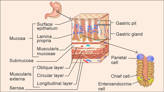
Mucosa refers to the inner lining of the stomach. Large, wrinkle-like folds called rugae lie in the mucosa when the stomach is empty. These flatten as the stomach fills and the stomach wall gets distended. It contains gastric glands, as well as pits. The mucosa is divided into the epithelial layer (non-ciliated simple columnar epithelium), lamina propria (layer of thin connective tissue), and muscularis mucosae (thin layer of smooth muscle).
Submucosa, which lies under the mucosa, is made up of connective tissue. Under the submucosa lies muscularis externa, which consists of layers of smooth muscle. Muscularis externa is composed of an outer longitudinal layer, a middle circular layer, and an inner oblique layer. It is the contraction of these muscle layers that helps in mixing, churning, and breaking down food. The outer layer that covers the stomach is called serosa. It is fused with visceral peritoneum, and is composed of simple squamous epithelium and areolar connective tissue.
Secretion of Gastric Juices
On the surface of the mucosa, the simple columnar epithelial cells or the goblet cells secrete mucus. The mucosa also contains tubule-shaped gastric glands that secrete gastric juices and mucus. Gastric pits, which are narrow channels in the stomach that act as openings for the gastric glands, are also lined by the surface mucus cells.
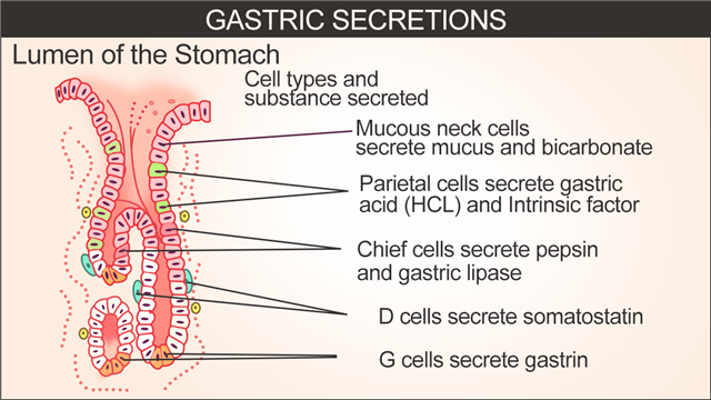
Gastric juices are secreted by special types of exocrine glands. These include neck mucous cells, chief cells, and parietal cells. The neck mucous cells secrete mucus that has a more neutral pH than that secreted by the cells at the surface of the stomach lining. The chief cells secrete pepsinogen, which is the inactive precursor to a proteolytic enzyme called pepsin. The conversion of pepsinogen to pepsin takes place in the lumen of the stomach, in the presence of gastric hydrochloric acid. The parietal cells secrete hydrochloric acid, which is a highly corrosive acid. It is hydrochloric acid that is responsible for the low pH (1-4) environment in the stomach. The parietal cells also secrete intrinsic factor, which is a glycoprotein that binds to vitamin B12 in the stomach and facilitates its absorption in the small intestine.
The G-cell, which is a type of hormone-producing cell in the lining of the gastric pits, produces and secretes a peptide hormone called gastrin either on receiving a signal from the vagus nerve, or due to the rise in the gastric pH of the stomach or distension of the stomach wall during a meal. Gastrin also promotes the production of hydrochloric acid by parietal cells.
The stomach receives parasympathetic nerve fibers from the vagus nerves and sympathetic fibers from the celiac ganglia. It must be noted that sensory vagal fibers play a role in gastric secretion. There are three phases of gastric secretion, which include the cephalic phase, gastric phase, and the intestinal phase. In the cephalic phase, parasympathetic reflexes are triggered due to the smell, sight, taste, or even thoughts about food. The gastric phase occurs with the entry of food into the stomach and the consequent rise in the pH and distension of the wall of the stomach. The intestinal phase commences as food enters the duodenum. It is characterized by the secretion of intestinal gastrin. During this phase, acid in the upper section of the small intestine triggers a sympathetic reflex, which in turn inhibits the secretion of gastric juice from the stomach wall.
Arteries of the Stomach
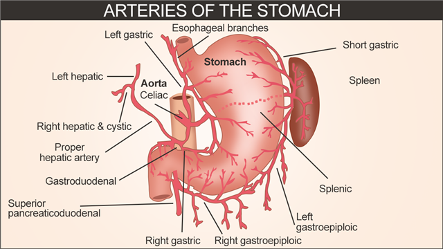
The stomach gets its supply of oxygenated blood from the branches of celiac artery. These branches include:
- Left gastric artery
- Right gastric artery
- Short gastric arteries
- Left gastroepiploic artery
- Right gastroepiploic artery
The left gastric artery is the narrowest branch of the celiac. It passes upward and runs to the left to reach the food pipe. Then, it runs down along the lesser curvature of the stomach. It provides blood to the upper right section of the stomach, as well as the lower section of the food pipe. The right gastric artery arises from the hepatic artery. Running to the left of the stomach’s lesser curvature and the pyloric end of the stomach, it joins the left gastric artery. This artery provides blood to the lower right section of the stomach. The short gastric arteries supply blood to the fundus. The left gastroepiploic and the right gastroepiploic arteries provide blood to the upper and the lower section of the greater curvature of stomach, respectively.
The veins of the stomach correspond to the aforementioned arteries. While the right and left gastric veins drain into the portal vein, the short gastric veins and the left gastroepiploic vein return blood into the splenic vein. The right gastroepiploic vein returns blood into the superior mesenteric vein.
On a concluding note, the main functions of the stomach include storage and mixing of food into chyme, and the passage of chyme into the duodenum. The cells in the stomach lining secrete gastric juices, with the maximum secretion taking place an hour after a meal. Problems can arise if the secretion of gastric juices or the wave-like muscle contractions (peristalsis) doesn’t take place properly.
