Nervous tissue, a component of nervous system, is made up of many neurons and supportive cells, called neuroglia. The main function of nervous tissue is to perceive stimuli and generate nerve impulses to various organs of the body. Let’s get to know its structure and functions in detail.
Did You Know?
The longest neuron in your body extends from your big toe all the way up to the base of your neck, making it almost as tall as you!
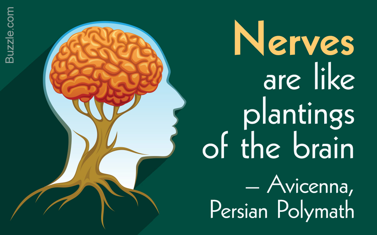
The human brain is the most awe-inspiring and fascinating organ in the human body. Every memory you have and every emotion you will ever experience is thanks to this remarkable result of evolution. The nervous system is essentially the powerhouse of your brain, comprising the nervous tissue. It is classified into two parts: (1) The CNS or Central Nervous System that encompasses decision-making centers of our body — namely the brain and spinal cord; and (2) the PNS or Peripheral Nervous System that consists of the remaining nervous tissues in our body.
Structure of a Neuron
In general, a neuron has three basic parts: (1) Cell body; (2) Axon; and (3) Dendrites.

1. Cell Body
The major part of the cell enclosed by a plasma membrane is the cell body, also known as the soma. It mainly comprises the nucleus and cytoplasm. Inside the cytoplasm, granules (Nissl bodies), mitochondria, golgi complex, lysosomes, and other cell organelles are present.
Function:
- Like in any other cell, the cell body is responsible for controlling metabolic activities.
- It powers the neuron by synthesizing energy and is in charge of the neuron’s growth and repair.
2. Axon
It is an elongated structure protruding away from the cell body and is severely branched at the end.
» Myelin Sheath: The axon is covered with a white fatty layer known as the myelin sheath.
Function:
- It serves two major functions — protecting and insulating the axon and accelerating the electrical signals during transmission.
» Neurilemma: The myelin sheath layer has a cellular covering known as the neurilemma or the Schwann cell sheath.
Function:
- The neurilemma is essential for regeneration of nerves. It is present only in the peripheral nervous system. In the central nervous system, neurilemma is absent, thus nerves here are incapable of regeneration.
» Nodes of Ranvier: The medullary sheath is not a continuous layer on the axon; it has joints, or node-type interruptions known as the nodes of Ranvier.
Function:
- They play a very important role in transmitting impulses. For any electrical signal to pass, there needs to be a potential difference between two given points. Nodes of Ranvier are these points, where the axon polarizes and depolarizes spontaneously, hence developing potential difference and transmitting the impulse.
- These nodes are also needed by the neuron for nutrition intake and waste disposal.
» Axon terminal: The axon branches out at the end, with small bulb-like endings, also known as terminal buttons. These branches are collectively known as the axon terminal.
Function:
- The axon terminal is responsible for transmitting impulses from one neuron to another. The terminal buttons produce neurotransmitters — a chemical essential for the transmission.
3. Dendrites
Numerous short-branched structures emerging from the soma are called dendrites. They are often covered with small, branched projections known as dendritic spines.
- Function of a Dendrite: Dendrites are the receptors of a neuron that receive electrical signals from other neurons.
- Function of Dendritic Spines: They increase the surface area of the dendrite vastly, thus helping in receiving impulses from other axons.
Synapse: Any two neurons are connected together at the synapse. This is where the electrical signal jumps from the axon terminal of one neuron to the dendrites of another neuron. Neurons aren’t actually connected; the axon and dendrites are merely close enough for the signal transmission to take place.
Types of Neurons
By Structure
» Unipolar: These neurons consist of one dendrite and one axon. The structure is such that a single projection emerges from the soma, splitting into two projections with one end having the axon terminal and the other having the dendrites. Such neurons are usually sensory neurons.
» Bipolar: Bipolar neurons have the most simplistic structure. The soma is located centrally, with the dendrites and axon protruding from opposite ends. These neurons are also mostly sensory neurons.
» Multipolar: Maximum neurons fall under this category, where the soma has multiple dendrites and a singular axon. These neurons are multifunctional and are found throughout our body.

By Functionality
» Afferent/Sensory Neurons: As the name suggests, these neurons are responsible for the transmission of impulses from the sensory organs to the spinal cord and the brain.
» Efferent/Motor Neurons/Motoneurons: They transmit impulses from the brain or spinal cord to the specific organ or muscle.
» Interneurons/Relay/Connector Neurons: These neurons transmit impulses between the above two types. They are mostly present within the brain or spinal cord.
Neuroglia
Neuroglia or glial cells are protective and supportive structures of the nervous tissue. They are found in bunches surrounding the neurons and have the ability to regenerate in case of injury.
Function: Neuroglia provide nutrition and immune protection to the neurons. In addition, they are responsible for the formation of myelin sheath and maintaining homeostasis inside the neurons. Some of the forms of neuroglia are astrocytes (provide metabolic support to nervous tissue), oligodendrocytes (support axons), and microglia (repair the damage of neurons), etc.
The nervous tissues of the peripheral nervous system are responsible for collecting signals from the organs and transmitting them to the central nervous system. Overall, the nervous system regulates and controls various functions of the body, such as memory, emotion, reasoning, and muscle contraction.


