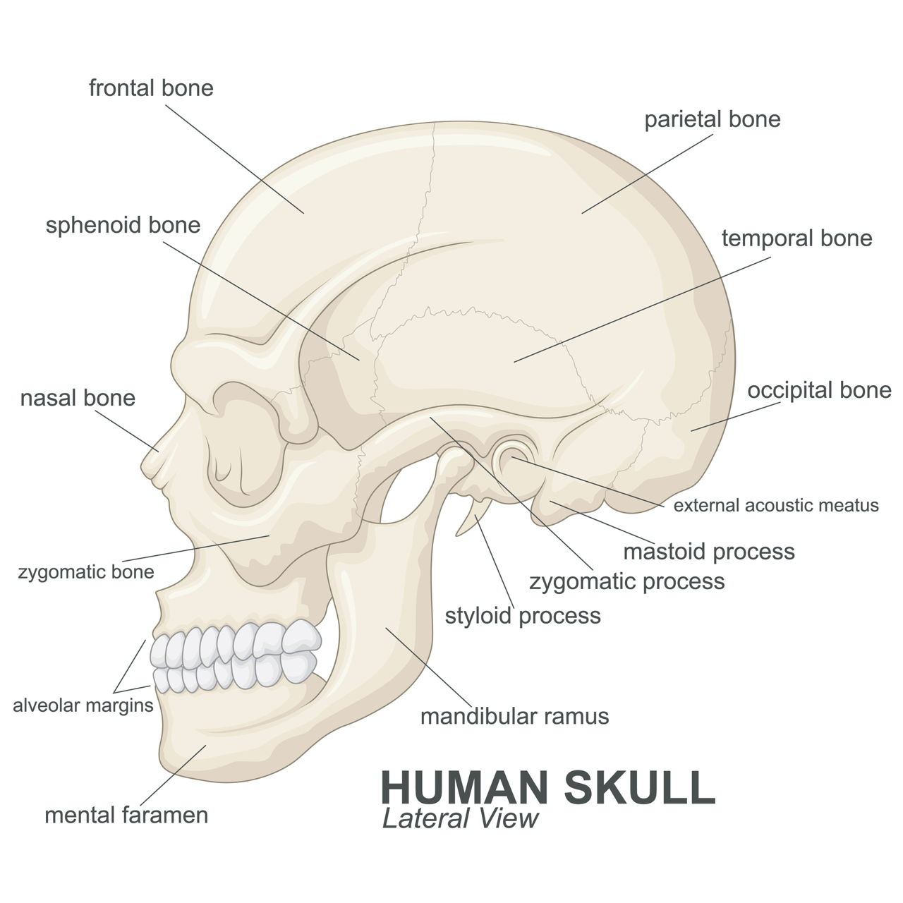
Certain muscles in the face are responsible for jaw movement, especially the mandible, which is the only movable joint in the skull. The muscles of mastication are paired on either side of the face.
Mastication is the first step of digestion of food in the human body. It is the process of chewing, crushing, and grinding the food with teeth with the help of enzymes in saliva. There are certain muscles, which assist in this process. These are the mastication muscles.
Anatomy of the Muscles of Mastication
In the human body, the lower jaw (also known as mandible) is connected to the temporal bone of the skull, via a complex joint known as temporomandibular joint. The mastication muscles start from skull to the mandible, allowing the flexible jaw movements when chewing.
There are four types of these muscles, which have different functions. These are discussed below.
| Muscle | Location | Function |
|---|---|---|
| Masseter Muscle | It consists of two overlapping muscles. The superficial head originates from the lower border of the zygomatic arch (cheek bone) of the skull. The deep head of the muscle rises from the inner surface of the zygomatic arch. Both the heads end into the ramus and the body of the mandible. The broad fibers of these muscles extend from second molar location on mandible to ramus. | These muscles are superficial, intermediate, and deep. They get the nerve supply from the masseteric nerve, and blood supply from maxillary artery (a terminal branch from external carotid artery). Masseter muscles are responsible for elevation of the mandible (when chewing/swallowing/talking), lateral movement of mandible (to effectively chew and grind food), and retraction of the mandible and unilateral chewing. It also is the strongest muscle in the human body. |
| Temporalis Muscle | It is a large fan-shaped muscle covering the temporal area of the skull. It starts from the parietal skull bone, and ends into the coronoid process of the mandible. | The temporalis muscle receives blood supply from anterior, posterior, and superficial temporal arteries, and receive nerve supply from temporal nerves. The main functions of these muscles include elevation and retraction of the mandible, and grinding food between the molars. |
| Lateral Pterygoid Muscle | It has two heads: inferior and the superior head. The former starts from the pterygoid process, and gets inserted into the mandible. The latter starts from the sphenoid bone, and gets inserted into the articular disc. | Lateral pterygoid muscles get the nerve supply from the pterygoid branch of trigeminal nerve. The arterial supply comes from the pterygoid branch of maxillary artery. The function of this muscle is to pull down the mandible, and protrude it in the forward direction to aid in the opening of jaw. |
| Medical Pterygoid Muscle | It is a thick muscle starting from the lateral pterygoid plate and maxillary tuberosity. It inserts itself into the medial angle of the mandible. | These muscles get their blood supply from the pterygoid branch of maxillary artery. Its main functions are elevating the mandible, closing the jaw, and moving it sideways. |
Accessory Muscles
| Muscle | Location | Function |
|---|---|---|
| Buccinator Muscle | This accessory muscle starts from the buccal plates of the bone, having sockets of the upper and lower three molars and pterygomandibular ligament. The buccinator muscle is found between the maxilla, and the mandible of the cheek bone. Upper fibers of this muscle merge into upper lips, lower fibers insert into lower lips, and middle fibers end at the opening of the mouth. | Buccinator muscles receive blood supply from facial arteries, and nerve supply from buccal branch of facial nerves. The main function of these muscles is to prevent accumulation of excess food in the vestibule of mouth. |
| Digastric Muscle (Anterior Belly) | It starts from the digastric fossa of the lower part of the mandible on either side of symphysis menti, and inserts itself into the intermediate tendon, which is connected to the hyoid bone. | Digastric muscle gets its nerve supply from the anterior division of mandibular branch of trigeminal nerve. Its main function is to depress the mandible. |
| Mylohyoid Muscle | It starts from the mylohyoid line of the internal mandible, and inserts itself into the hyoid bone from below, and into the chin from above. Mylohyoid muscle forms the floor of the mouth. | It receives nerve supply from the inferior alveolar branch of mandibular nerve (that starts before entering the inferior alveolar canal). These muscles receive blood supply from both lingual and facial arteries, and their main function is to support the floor of the mouth during depression of mandible (like swallowing) |
| Geniohyoid Muscle | It starts from the inferior genial tubercle, and is inserted into the hyoid bone. | Geniohyoid muscle receives its blood supply from lingual artery, and nerve supply from hypolossal nerve. The main function of this muscle is to depress the mandible when swallowing and talking. |
| Orbicularis Oris Muscle | It is divided into intrinsic and extrinsic parts. The former is a thin sheet of muscle starting from inferior and superior incisivus, and is inserted into the angle of the mouth. The latter one of the orbicularis oris muscle is formed by the elevator and depressor muscles of lips. This is also inserted into the angle of mouth. | Orbicularis oris muscles receive blood supply from facial arteries and nerve supply from facial nerves (which are one of the cranial nerves). The major function of this muscle is to aid in opening and shutting of the mouth, thereby controlling the movements of lips and cheeks. |
These muscles are an integral part of our human body systems, as they are responsible for our facial expressions and talking. Any injury inflicted on them can lead to temporomandibular joint disorders. Regular jaw exercises help in maintaining healthy jaw muscles.


