The human kidneys house millions of tiny filtration units called nephrons, which enable our body to retain the vital nutrients, and excrete the unwanted or excess molecules as well as metabolic wastes from the body. In addition, they also play an important role in maintaining the water balance of our body.
Quick Facts
Size of an adult kidney:
Length: 11-12 cm
Width: 5.0-7.5 cm
Weight of an adult kidney:
Males: 125-170 g
Females: 115-155 g
Located in the abdominal cavity, kidneys are the most efficient filters. They are an important component of the human excretory system, and help the body retain essential molecules and get rid of the unwanted ones. They play a vital role in maintaining the composition and volume of body fluids.
Cross-section of a Human Kidney
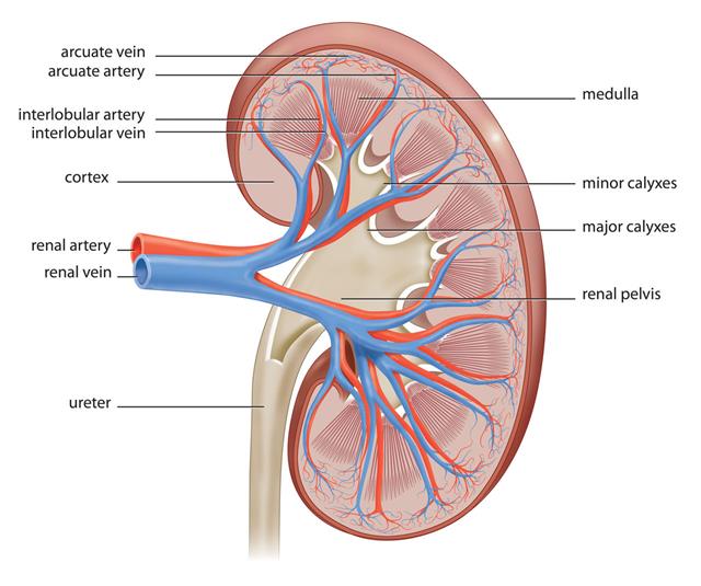
- The vital structural components of a kidney are enclosed in a smooth but tough fibrous capsule called renal capsule.
- Inside this capsule, two distinct regions can be observed: a pale outer region called renal cortex, and a dark inner portion called renal medulla.
- The renal medulla comprises a set of 8-18 conical structures called renal pyramids that are surrounded by the cortex. Portions of the cortex between two adjacent pyramids are termed as renal columns.
- Spread in these pyramids and the cortex, are the functional units callednephrons. The actual filtration of blood occurs in the nephrons.
- The tips of the renal pyramids are called renal papilla, and are surrounded by cup-shaped drains called minor calyces (singular: calyx).
- The minor calyces converge to form two or three larger drains called major calyces, which eventually converge to a structure called the renal pelvis. The renal pelvis is connected to the ureter.
- The blood supply to all these structures occurs through the branches and sub-branches of the renal artery called interlobular arteries and arcuate arteries respectively. The interlobular arteries supply blood to the borders of the cortex and medulla, whereas the arcuate arteries diverge to form afferent arterioles that carry blood to the nephrons for filtration.
- The filtered blood is ultimately collected through venules and sub-branches called interlobular veins and arcuate veins which converge into the renal vein.
- The waste fluid or urine is collected in a common collecting duct of the nephrons and is emptied into the minor calyces. The minor calyces drain into the major calyces, which empty their contents into the renal pelvis. From here, it is carried by the ureter to the urinary bladder.
Structure of a Nephron

Each kidney contains more than 800,000 nephrons, each of which serves as the basic functional unit by performing three essential processes: filtration, reabsorption and secretion. The nephron essentially comprises renal corpuscle and renal tubule.
Renal corpuscle

- It is made up of a network of capillaries called glomerulus which arise from the afferent arteriole, and exit the renal corpuscle as efferent arteriole; and a cup-like sac called Bowman’s capsule that surrounds the glomerulus. The initial step of filtration occurs in the renal corpuscle.
- The walls of the glomerular capillary are composed of a three-layered filter that allows the filtration of small molecules, and is not permeable to large macromolecules like albumin and blood cells.
- A high pressure is created in the glomerular capillaries because the efferent arteriole has a smaller diameter as compared to the afferent arteriole. As a result of this high pressure, ions and water molecules are forced into the Bowman’s capsule, leaving behind concentrated blood containing blood cells and macromolecule.
- The filtrate thus extracted into the Bowman’s capsule is termed glomerular ultrafiltrate. It passes from the Bowman’s capsule into the renal tubule, while the concentrated blood flows out of the glomerulus through the efferent arterioles.
- The entire blood volume gets filtered about 20-25 times per day through such ultrafiltration process.
Renal tubule
- It is a specialized tubular structure made up of proximal convoluted tubule, a U-shaped tube called Loop of Henle, and a distal convoluted tubule. These three tubular components are selectively permeable and only allow specific molecules to pass through them. The renal tubule is surrounded by capillaries called peritubular capillaries that arise from the efferent arterioles.
- The renal tubule is the site for reabsorption and secretion. The substances essential for the body are reabsorbed from the tubules into the peritubular capillaries, and the unwanted or toxic molecules are secreted into the lumen of the renal tubule.
- Water, sodium and potassium ions, urea, phosphate, citrate, as well as organic molecules like glucose and amino acids are reabsorbed from the proximal convoluted tubule. In addition, it is the site for formation of ammonium and also involves the secretion of excess medicines from blood.
- This filtrate then enters into the descending loop of Henle, which lies in the medulla of the kidney. Here, reabsroption of water from the filtrate to the tissues takes place. This water is transferred by the cells to the capillaries surrounding them.
- The filtrate then travels through the ascending loop of Henle which is impermeable to water. Hence, only ions diffuse out into the surrounding cells, which subsequently pass them into the surrounding capillaries.
- While passing through the distal convoluted tubule, the surrounding tissues further facilitate the exchange of water and ions from the filtrate to the capillaries. They also absorb the excess potassium and hydrogen ions from the capillaries and secrete them into the filtrate.
- The filtrate from several nephrons is then collected into the common collecting duct which empties into the minor calyces, and is subsequently collected into the bladder as urine.
The kidneys play a vital role in water and ion homeostasis, and contain fascinating biological filtration assembly. Any defects or discrepancies in the normal functioning of the kidneys takes a toll on the many cellular and physiological processes of the body.


