Wondering what is the structure of the human ear, and how it performs the function of hearing? Look no further, this Bodytomy article gives you a labeled human ear diagram and also explains the functions of its different components.
The human body is like a big machine, and various processes take place inside it. With the help of the various organs and tissues, it carries out some of the most marvelous tasks, that are no less than a miracle! One such organ is the ear that helps us in the process of hearing and balancing. The sound waves entering the ear get converted into electric impulses for the brain to understand and interpret.
Let us take a look at the human ear structure with the help of a diagram, and understand its functions a little more closely.
The Structure of Human Ear

- Helix: It is the prominent outer rim of the external ear.
- Antihelix: It is the cartilage curve that is situated parallel to the helix.
- Crus of the Helix: It is the landmark of the outer ear, situated right above the pointy protrusion known as the tragus.
- Auditory Ossicles: The three small bones in the middle ear, called malleus, stapes, and incus, are connected. These bones together are called the auditory ossicles, and their purpose is to let the sound that strikes the eardrum, further into the inner ear.
- Oval Window: Oval window is the opening covered by a thin membrane, which connects the middle ear to the inner ear.
- External Auditory Canal: External auditory canal or ear canal, is the channel from which the sound enters from the outside ear to the eardrum.
- Eardrum/Tympanic Membrane: It is the thin membrane located between the external ear canal and the middle ear.
- Cochlea: Cochlea is tiny conical structure situated in the inner ear that resembles a snail shell. It is responsible for converting sound vibrations into nerve signals that are sent to the brain.
- Eustachian Tube: It is a tube which connects the middle ear cavity and the pharynx. It plays a crucial role in ensuring that the air pressure on each side of the eardrum is even.
- Semicircular Canals: The semicircular canals are located in the inner ear and constitute of three tiny tubes that are filled with fluid. They play a crucial role when it comes to balancing the body.
- Vestibulocochlear Nerve: It is nerve that transmits messages regarding sound and balance, from the inner ear to the brain.
How Do We Hear?
The sound waves travel first through the ear canal and vibrate the eardrum. Before the sound waves enter the inner ear, the total pressure must be amplified. The ossicles in the middle ear do the job of amplification. Then the cochlea in the inner ear conducts the sound through a fluid. Cochlea is a complex part of the ear, which takes the physical vibrations caused by the sound waves and translates them into electrical information for the brain to recognize as sound. The organ of corti which contains thousands of tiny hair cells, when moved, sends electrical impulses through the cochlear nerve. The cochlear nerve then passes these impulses to the cerebral cortex and the brain interprets them, and we get to hear.
Quite interesting, wasn’t it? How the ear consists of so many tiny little components―that are almost nonexistent to the common man―that together make the functioning of this organ possible. We are sure that the aforementioned information must have added to your knowledge about the human body.
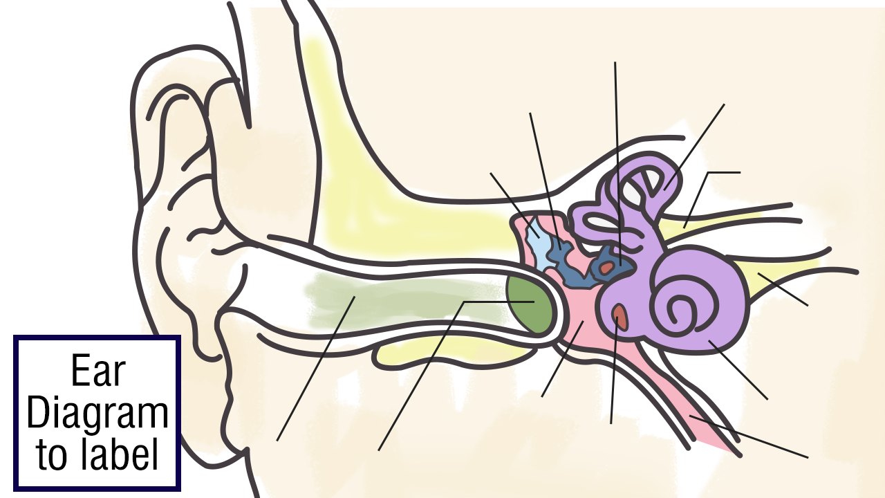
Here is a blank human ear diagram for you to label, so that you can memorize the different parts of this vitally necessary organ, for good.