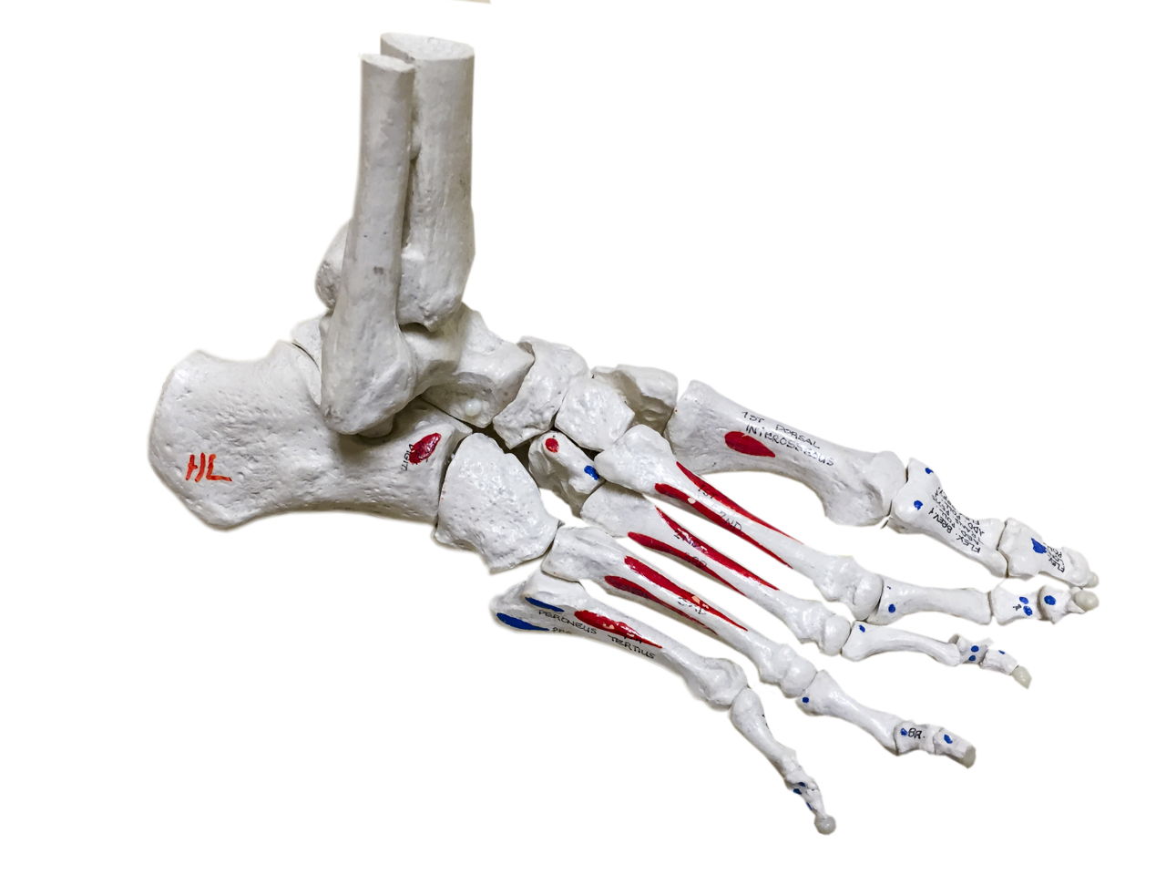
There are numerous bones located in the foot. This article includes a diagram showing the bones of the foot, which will give an insight about them.
Bones, muscles, ligaments, and tendons make up the foot. The bones of the foot are divided into anterior region, posterior region, dorsal region, plantar region, distal region, proximal region, medial region, and lateral region. It is important to note, when we say a joint, it is not only about two bones coming together, the muscles, ligaments, and tendons are also a part of it. Likewise, along with the bones, there are muscles, ligaments, and tendons in the foot. All of us are aware about how many bones are present in the human body. Out of 206 bones present in the body, 28 bones are located in the human foot alone. This includes 2 sesamoid bones of the foot. The foot is made up of 33 joints. In this article, to make it easier to undertand, the bones of the foot will be divided into tarsal bones, metatarsal bones, and phalanges. This diagram of the foot will prove beneficial in understanding the bones of the foot better.
When one looks at the anatomy of the foot, they would realize that the foot has a complex mechanical and structural architecture. The ankle joint is the shock absorber of the foot. Apart from 28 bones, 33 joints, muscles, ligaments, and about 100 foot tendons make the foot. The diagram of bones in the ankle and foot is given below:

Tarsal Bones
The tarsal bones in the foot are located amongst tibia, metatarsal bones, and fibula. There are in all 7 bones, which fall under tarsal bones category. They are:
- Calcaneus or Calcaneum: To explain the term in layman’s language, it is the heel bone in the skeletal system. It is situated between talus and cuboid.
- Talus: This bone is sandwiched between tibia at the top, fibula around the sides, and calcaneus below.
- Navicular: This bone derives its name from its shape. It resembles a boat. It is surrounded by talus and the cuneiform bones.
- Medial Cuneiform: When one takes a top view of the foot, the bone located in the mid of the foot is the medial cuneiform.
- Intermediate Cuneiform: The shape of this foot bone is similar to that of a wedge. It is stuck between the other two cuneiform bones.
- Lateral Cuneiform: This bone is located in the front row of the tarsal bones. The other name of this bone is external cuneiform.
- Cuboid: The name of this bone is also derived from the way it looks. It is surrounded by metatarsal bones and calcaneus and the navicular bones.
Metatarsal Bones
On one end of each of the metatarsal bones are the tarsal bones and on the other end are the phalanges. In other words, they are situated exactly between the tarsal bones and phalanges. There are in total 5 bones, which fall under metatarsal bones. These bones in the human body system are known as:
- First Metatarsal Bone
- Second Metatarsal Bone
- Third Metatarsal Bone
- Fourth Metatarsal Bone
- Fifth Metatarsal Bone
The second metatarsal bone is the longest of all the metatarsals, while the first metatarsal is the shortest, yet thickest of the metatarsal bones. Injuries are common to the second and fifth metatarsal bones among athletes.
Phalanges
The bones situated in the toe region are known as phalanges. Every human foot has 14 phalanges. Other than the big toe, the structure of all the toes is the same with three phalanges, namely proximal phalanx, middle phalanx, and distal phalanx. The big toe on the other hand only has two phalanges, namely proximal phalanx and distal phalanx.
- Proximal Phalanx: These phalanges are situated closer to the foot, hence the name proximal phalanx.
- Middle Phalanx: These bones in the foot are situated further ahead from proximal phalanx. The big toe does not have a middle phalanx.
- Distal Phalanx: The tip of the toes are the distal phalanx. When one takes a look at the foot bones diagram, one would observe that the nails are situated on the distal phalanx.
The foot is the weight bearing part of the body, hence, care should be taken not to injure the foot, to avoid inconvenience of any kind.


